|
Definition of Ganglion Cysts:
Ganglion cysts are the most common type of benign tumors that form on the hand or the wrist.
Ganglion cysts come from nearby joints or tendon sheaths. They are sphere-, oval-, or sometimes sack-shaped formations with a
stem that is bound to either a joint capsule or a tendon sheath. They are filled with a more or less clear fluid or gelatinous
substance. When examined, they appear as a small water sac or taut, elastic swelling. Ganglion cysts can occur on all joints
and tendon sheaths of the hand and wrist, though they tend to be more common in certain locations:
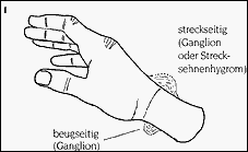
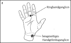
- Transition between the wrist and carpus - extensor-side, radial-side
- Transition between the wrist and carpus - flexor-side, radial-side
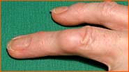
- At the height of the flexor-side of the finger basal joints as so-called annular pulley ganglion cysts
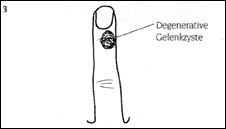
- At the height of the flexor-side of the finger basal joints as so-called degenerative joint cysts within the context of degenerative joint alterations (Heberden's rheumatism)
- On the back of the hand, arising from extensor tendon sheaths, also know as hygroma
Causes of Ganglion Cysts
Ganglion cysts are always benign!
A clear cause cannot be provided though there is discussion of links with injuries, wear and tear, and most often a constitutional,
local weak point of the joint capsule or tendon sheath. Pressure within a joint or tendon sheath can cause fluid to press through
these weak points and thereby creates a sac-like cyst full of fluid.
Signs and Symptoms of Ganglion Cysts (Illustrations 1 & 2)
Ailments depend on the location and size of the cysts.
- Ganglion Cysts of the Wrist
- Size: variable, diameter rarely larger than 2 cm
- Pain: independent of size (even small cysts can hurt); pain is rarely the first sign of an inconspicuous ganglion cyst.
- Limitation of Maneuverability: depends on level of pain and the size of the ganglion cyst, but seldom occurs.
- Cosmetic nuisance: depends on the size and position of the cyst.
- Annular Pulley Ganglion
- Size: variable, diameter rarely larger than 1 cm.
- Pain: typical pressure pain when gripping steering wheel, door knobs etc.
- Limitation of Maneuverability: depends on level of pain and the size of the ganglion cyst, but seldom occurs.
- Degenerative joint cysts
- Size: variable, as a rule 0.5 - 1.0 cm
- Pain: independent of size, usually superimposed by pain from arthritis of the affected distal interphalangeal joint of the finger.
- Limitation of Maneuverability: usually caused by the arthritis
- Nail growth malformations: depending on the position of the cyst relative to the root of the finger nail.
- Extensor Tendon Hygroma
- Size: variable, changing, can grow considerably larger than ganglion cysts
- Composition: in contrast to ganglion cysts, these are soft and elastic
- Pain: independent of size, but seldom occurs
- Limitation of Maneuverability: seldom
- Cosmetic nuisance: depends on the size and position of the hygroma
Diagnosis of Ganglion Cysts
As a rule, the typical case history (anamnesis) and a clinical examination with the determination
of the described symptoms leads to a reliable diagnosis. The skin over the ganglion cyst is generally easy to move around while
the cyst itself is usually firmly connected to the tendon sheath or joint capsule and is not easily moved unless the stem is
very long.
The size of ganglion cysts can wax and wane. They can spontaneously shrink or disappear altogether.
A definitive diagnosis can only be made by puncturing the tumor and obtaining a sample of the fluid or through the surgical
removal and examination of tissue. A histological examination is necessary since other benign and very rare (!) malignant
tumors can also be similar in appearance to ganglion cysts.
In order to clarify whether or not injuries or alterations of the bone or joint exist before surgery, an x-ray examination
of the affected area and, if necessary, the healthy hand should be performed.
Treatment of Ganglion Cysts
- Non-surgical:
- Puncture: in approximately 50 % of the cases a reoccurrence should be expected!
- Immobilization (using a splint): pain is rarely relieved and the ganglion cyst is not likely to diminish in size.
- "Monitoring": no particular therapy, appropriate when pain and cosmetic nuisance are minimal.
- Surgical:
Surgery is appropriate in cases of bothersome pain and/or mobility limitations as well in cases of cosmetic
disturbance. Pain in the wrist area cannot always be attributed to the presence of ganglion cysts. Sometimes
other causes exist such as capsule-ligament instability. In such cases the pain does not diminish or disappear
through surgery. Even in cases of proper surgical and post-operative treatment techniques, patients must expect
a 10-20% chance of a reoccurrence of the ganglion in the same position.
Risks Associated with Surgery:
Overall very minimal!
- Blood and wound infection very rare
- Mobility limitation usually temporary
- Damage of healthy, neighboring tissue (nerves, tendons, vessels) is avoided through exact surgical techniques,
expert knowledge of anatomy, exsanguination providing a clear field of view, and the use of magnifying glasses.
Anesthesia:
There are various possibilities for ensuring that the patient is free of pain during the surgery.
These possibilities will be explained to you by the anesthesiologist. Gegenerative cysts in the area of the distal
interphalangeal joint are operated upon after the finger is numbed (Oberst's nerve block).
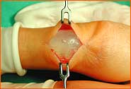
Technical Aspects of the Surgery:
Surgery of ganglion cysts is usually out-patient, that is, the patient can go home once the operation is completed.
1. General surgical preparation:
- Exsanguination:
In order to provide optimal viewing conditions and to reduce the risk of damage to healthy,
neighboring structures (nerves, vasculature, tendons) during hand surgery, so-called exsanguination is necessary.
The entire arm is wrapped in a rubber bandage which pushes the blood out of the arm. A cuff is then placed on the
arm to prevent blood from reentering the arm during the course of the surgery.
- Skin disinfection and sterile conditions:
In order to decrease the risk of infection, the skin is disinfected and the surgical field is covered with sterile cloth.
- Magnifying glasses:
The surgeon utilizes magnifying glasses in order to see and manipulate the hand structures as well as possible.
2. Surgery Sequence of Events:
- Incision
- Exposure of nerves and vessels of the subcutaneous layer
- Exposure of the tendon sheaths and tendons
- Exposure of the cyst except for the most inner layer
- Dissection of the cyst through to the stem which adheres to the tendon sheath or joint capsule
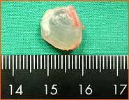
- Excision of the cyst, tissue sample sent for histological examination
- Smaller defects in the joint capsule or tendon sheath can be sutured while larger defects are left open and scarred over
- Final control of the sound condition of the visible structures
- Wound suture
- Sterile compression band applied
- In cases of ganglion cysts, immobilization is ensured for 14 days through wearing of a splint. In cases of a hygroma, annular pulley ganglion cysts and degenerative joint cysts, no immobilization is necessary
Post-operative treatment:
- The patient is allowed to return home after the surgery. Joints not in a cast should be moved but should not bear any weight.
- The hand operated upon should be more or less constantly elevated by stretching the hand above the head every half hour for 2-5 minutes.
While sitting or lying, hand should be placed on pillow level with the heart Goal: less secondary bleeding, less swelling, less pain, better healing!
- One day after surgery: Cast and soft tissue control (can also be performed by family physician)
- Five to seven days after the surgery: bandage change (can also be performed by family physician)
- Fourteen days after surgery: Removal of splint, end of immobilization period, bandage change and removal of
stitches (can also be performed by family physician).
- The bandage is no longer necessary the day after the stitches are removed. Begin of regular (3-4 times daily)
exercises in cold water (with addition of ice cubes). Cold reduces swelling and pain. Patients that cannot tolerate
cold should use lukewarm water.
- The scar treatment can begin five days after the removal of stitches. The scar should be massaged with a fatty
cream or salve 4-5 times daily so that the scar becomes softer, less painful, and more resistant to strain
("toughening" of the scar). Also beneficial is the tapping of the scar, for example with a soft brush.
- Physical and/or occupational therapy is usually not necessary except in cases of mobility limitations.
- The duration of the patient's incapacity to work is usually around three to four weeks.
Healing progression after the surgery:
As a general rule, patients can return to work three to four weeks after surgery of ganglion cysts of the wrist.
A decrease in pain caused by the cyst does not always occur (see above).
After the surgery, pain and mobility limitations depending on the type of movement and strain can continue for several weeks
and thereby influence the ability to return to work.
Pain usually disappears directly after surgery of extensor tendon hygroma and annular pulley ganglion cysts and patients can
resume all activities 2-3 weeks after surgery. Scar discomfort largely disappears after the first 6-8 weeks. After 3-6 months
patients no longer complain of pain. However, the scars final state is not reached until approximately 12 months after surgery
© Dr. Klaus Lowka
 back to top back to top
|
|
 |