|
||||||
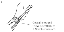
| Definition of de Quervain's Tendinitis: compartment. In the past it was known as "washer women's sprain" and is today named after the man that first described the condition, the Swiss surgeon Fritz de Quervain (1895). The extensor tendons run along the extensor-side of the forearm to the back of the hand through six compartments. This involves very strong structures (fibrous tissue). At times, the extensor tendons run through U-shaped bone canals. The extensor tendons are held in place by the transverse retaining ligament (extensor retinaculum), which wraps around the wrist like a watch (Illustration 1). 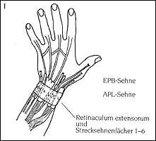 The first dorsal compartment lies over the radial malleolus (styloid process of radius) proximal to the base of the thumb. The large tendon of the abductor pollicis longus (APL) and the small extensor pollicis brevis (EPB) tendon pass through the first dorsal compartment (Illustration 2). 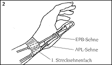 The APL tendon often has multiple splits in the lengthwise direction. During muscle contraction it spreads the thumb in the saddle joint from the hand. The EPB tendon extends the thumb in its base joint. Within the first dorsal compartment the EPB tendon runs completely or partially in a separate compartment in many cases. A non-thorough surgical technique may result in an incomplete splitting of the first dorsal compartment. Both tendons (APL and EPB) aid in spreading the thumb away from the rest of the hand and straightening the thumb for grasping. They also play a minor role in the stabilization and mobilization of the wrist. The condition of de Quervain's tendinitis involves the painful inflammation of the tendons and their lubricating lining in the first dorsal compartment. The swelling of the tendon sheath tissue and, as the case may be, the tendon tissue creates constricted conditions within the dorsal compartment. This constriction causes pain as the tendons attempt to glide through the compartment and can also lead to a cracking or creaking sensation (crepitation) that can be both felt and heard. The inflammation can also lead to adhesion between the tendons and the tendon sheaths. Causes of de Quervain's Tendinitis This condition typically arises in individuals between the ages of 30 and 50 and affects women eight times more frequently than men. This condition can be evoked by changes in the structure of the first dorsal compartment (e.g. after distal or radial fractures near the wrist) or by the swelling of the tendons or parts of the dorsal compartment. Repeated injuries, overuse and inflammatory disease are causes that can trigger a predisposition for the condition, but in most cases the causes are not readily known. Individuals that perform tasks that require the repetitive side-to-side movement of the wrist while simultaneously providing stabilization (hammering, utilization of ski poles), can be predisposed to this condition. Signs and Symptoms of de Quervain's Tendinitis Pain develops in the area of the first dorsal compartment temporarily, recurrently or "overnight" depending on the extent of the inflammation. The pain can radiate into the thumb and the extensor and radial side of the wrist of the hand as well as the extensor and radial side of the fore arm (dispersing over the superficial, sensitive radial nerve branches). Pain can increase depending on the type of movement and the amount of weight especially during a firm grip, finger tip grip and rotational movements. A more or less pronounced swelling is evident over the first dorsal compartment and one can trigger evident pain through application of pressure and tapping of the area. In contrast to trigger finger (stenosing tenosynovitis), a so-called "dorsal snapping" is seldom observed in cases of de Quervain's tendinitis. A classic phenomenon is a positive Finkelstein test: the thumb is bent into the palm as tightly as possible and a fist is made with the fingers over the thumb. The wrist is then moved suddenly, quickly and firmly towards the ulna (little finger side of the hand) (Illustration 3). When the test is positive, the patient experiences a very sharp, sometimes almost electrical pain in the area of the first dorsal compartment with distal radiation. Simultaneously, a cracking (crepitation, snapping) can also often be felt and/or heard. 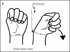 Diagnosis of de Quervain's Tendinitis As a rule, the typical case history (anamnesis) and a clinical examination with the determination of the described symptoms leads to a reliable diagnosis. In order to rule out any bone-related causes of the ailments, an x-ray examination should be performed on the affected hand, with comparison of the healthy hand if necessary. The x-ray examination should always precede any surgical action. Treatment of de Quervain's Tendinitis
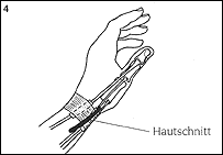
There are various possibilities for ensuring that the patient is free of pain during the surgery. These possibilities will be explained to you by the anesthesiologist. Technical Aspects of the Surgery Surgery for de Quervain's tendinitis is usually out-patient, that is, the patient can go home once the operation is completed. 1. General surgical preparation:
Pain as a result of the incision is typically minimal and most patients do not require pain medication. The pain symptoms typical of the condition disappear after surgery while the radiating pain diminishes after a few days. In rare cases, the patient still notices the rubbing together of the tendons, though this sensation disappears after a few weeks. Scar discomfort largely disappears after the first 6-8 weeks. After 3-6 months patients no longer complain of pain. However, the scars final state is not reached until approximately 12 months after surgery. © Dr. Klaus Lowka |
||||
| Home | About Dr. Lowka | Advanced Education/Publications | The Hand Hand Surgery Diagnosis and Operations | Clinical Pictures in Hand Surgery | Areas of Emphasis in Hand Surgery Post-operative Treatment | Center for Diagnosis and Out-patient Surgery Contact Us | Legal Information |
||||